Extra-Large Area 2D/3D Imaging

Mult-Beam Scanning Electron Microscopy is an enabling technology

Many scientific questions need submicron image resolution over large sample areas. This is impractical with conventional SEMs (months or years acquisition time). Multi-Beam Scanning Electron Microscopes (MultiSEM) can resolve these limitations.

MultiSEM uses multiple electron beams (green: illumination path) and detectors in parallel. A finely tuned detection path (red) collects a large yield of secondary electrons (SE) for imaging. Each beam carries out a synchronized scanning routine at one sample position, resulting in a single sub-image. The full image is formed by merging all image tiles. A parallel computer setup is used for fast data recording, ensuring high total imaging speed. Image acquisition and workflow control are fully separated in the MultiSEM system.

More information on the instrumentation is available on the vendor websites, e.g. Carl Zeiss Microscopy (Instrumentation / Technology). NanoWorldMaps works in improving the availability of the Ultra-Fast 2D/3D Imaging technology in Europe.

Multi-beam Scanning Electron Microscopy technology

Leading the revolution in high-throughput multimodal imaging at nanometre scale

Multi-SEM is the platform technology to capture square centimetre-sized structures on a nanometer scale. It creates an ultra-dense map of the nanoworld. Artificial Intelligence (AI) methods can then be used to analyse the functionality of the sampled system or to search for and locate Points of Interest. This approach is a very good basis for multimodal analysis of the identified Points of Interest. Different types of analytical technologies such as Raman Spectroscopy (RAMAN) or Secondary Ion Mass Spectrometry (SIMS) can approach the identified points of interest and investigate them in detail.

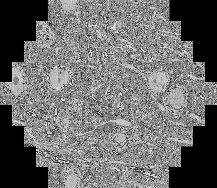
Overview image of a 50 nm thick mouse brain section acquired by MultiSEM

Case studies

NanoWorldMaps members are working on a series of case studies to show the potential of Extra-large 2D/3D imaging:

  • 3D tissue/organ imaging – hypothalamus: Image registration for 3D multi-beam SEM and multimodal data.

  • 3D tissue/organ imaging – bone: Bone is a material with irregular structural features at many size scales. Combination of Multi-beam SEM with e.g. RAMAN, SIMS or XRM

  • Membranes for molecular selective ultrafiltration: A non-flat, smooth material, image processing challenges similar to those for battery electrodes.

  • Nanoplastics from environmental sampling: Non-conductive samples, loose particles distributed over large area.
  • Lithium-ion battery electrodes and electrolyte: Small, high aspect ratio feature recognition, air sensitive. AI for recognition of faults and cracks.
  • Corrosion and cracking of metals: Used in nuclear power plants. Small, rare/sparse feature recognition.
  • Defects in electronic devices: Circuit interconnection include interfaces, crack and void detection.
Sample preparation by microtome slicing on tape sample holders

Publications

Extra-Large 2D/3D Imaging, MultiSEM and combined multimodal approaches (RAMAN, SIMS) attracted significant scientific interest. Some publications:

  • Alexander Shapson-Coe, Michał Januszewski, Daniel R. Berger, Art Pope, Yuelong Wu, Tim Blakely, Richard L. Schalek, Peter H. Li, Shuohong Wang, Jeremy MaitinShepard, Neha Karlupia, Sven Dorkenwald, Evelina Sjostedt, Laramie Leavitt, Dongil Lee, Luke Bailey, Angerica Fitzmaurice, Rohin Kar, Benjamin Field, Hank Wu, Julian Wagner-Carena, David Aley, Joanna Lau, Zudi Lin, Donglai Wei, Hanspeter Pfister, Adi Peleg, Viren Jain, Jeff W. Lichtman (2021): A connectomic study of a petascale fragment of human cerebral cortex, Preprint on bioRxiv, https://www.biorxiv.org/content/10.1101/2021.05.29.446289v4

  • Anja Günther, Karin Dedek, Silke Haverkamp, Stephan Irsen, Kevin L. Briggman, Henrik Mouritsen (2021): Double Cones and the Diverse Connectivity of Photoreceptors and Bipolar Cells in an Avian Retina, Journal of Neuroscience 9 June 2021, 41 (23) 5015-5028; DOI: 10.1523/JNEUROSCI.2495-20.2021 https://www.jneurosci.org/content/41/23/5015

  • Hayworth, K.J., Peale, D., Januszewski, M. et al. (2020): Gas cluster ion beam SEM for imaging of large tissue samples with 10 nm isotropic resolution. Nat Methods 17, 68–71. https://doi.org/10.1038/s41592-019-0641-2

  • David Zhang, Gooitzen van der Wal, Phil Miller, David Stoker, Erik Matlin, Naveen Marri, Gary Gan, Joe Zhang, Jane Asmuth, Sek Chai, David Weaver, Michael Piacentino, Scott Silverman, Michael DiBattista, Robert Chivas, Christopher G Ferri, David Taylor, Jordan Furlong, Thomas Harper, Dustin Kobs (2019): “Fast, Full Chip Image Stitching of Nanoscale Integrated Circuits,” Proceedings GomacTech, https://pdfs.semanticscholar.org/1fe4/834b9841d82863a20cdc0d99ad02f255a667.pdf

  • Anton D. Nathanson, Lucy Ngo, Tomasz Garbowski, Abhilash Srikantha, Christian Wojek, Dirk Zeidler, Melissa L. Knothe Tate (2019): “Work Flows for Cellular Epidemiology, From Conception to Translation,” bioRxiv 548412; doi: https://doi.org/10.1101/548412

  • Eberle AL and Zeidler D (2018): “Multi-Beam Scanning Electron Microscopy for High-Throughput Imaging in Connectomics Research,” Front. Neuroanat. 12:112, doi:10.3389/fnana.2018.00112

  • Haehn, D.; Hoffer, J.; Matejek, B.; Suissa-Peleg, A.; Al-Awami, A.K.; Kamentsky, L.; Gonda, F.; Meng, E.; Zhang, W.; Schalek, R.; Wilson, A.; Parag, T.; Beyer, J.; Kaynig, V.; Jones, T.R.; Tompkin, J.; Hadwiger, M.; Lichtman, J.W.; Pfister, H. (2017): Scalable Interactive Visualization for Connectomics. Informatics, 2017, 4, 29.

  • M. L. Knothe Tate, D. Zeidler, A. F. Pereira, D. Hageman, T. Garbowski, S. Mishra, L. Gardner, and U. R. Knothe (2016): “Organ-to-Cell-Scale Health Assessment Using Geographical Information System Approaches with Multibeam Scanning Electron Microscopy,” Advanced Healthcare Materials, vol. 5, no. 13, pp. 1581–1587, Jul. 2016.

  • S. Mikula (2016): “Progress Towards Mammalian Whole-Brain Cellular Connectomics,” Frontiers in Neuroanatomy, vol. 10, Jun. 2016.

  • J. R. Michael, C. Y. Nakakura, T. Garbowski, A. L. Eberle, T. Kemen, and D. Zeidler (2015): “High-Throughput SEM via Multi-Beam SEM: Applications in Materials Science,” in Proceedings of Microscopy & Microanalysis 2015, Portland, Oregon, 2015, vol. 21 (Suppl 3), pp. 697–698.

  • A. L. Eberle, S. Mikula, R. Schalek, J. W. Lichtman, M. L. Knothe Tate, and D. Zeidler (2015): “High-resolution, high-throughput imaging with a multibeam scanning electron microscope,” J Microsc, Jan. 2015.

  • A. L. Eberle, R. Schalek, J. W. Lichtman, M. Malloy, B. Thiel, and D. Zeidler (2015): “Multiple-Beam Scanning Electron Microscopy,” Microscopy Today, vol. 23, no. 2, pp. 12–19, Mar. 2015.

  • S. Mikula and W. Denk (2015): “High-resolution whole-brain staining for electron microscopic circuit reconstruction,” Nat. Methods, Apr. 2015.